Dental CBCT Scan: Improving Diagnosis
A Dental CBCT Scan in Dubai provides detailed three-dimensional images of the teeth, jawbone, roots, and surrounding oral structures. This additional level of imaging can help dentists evaluate areas that may be difficult to understand through a standard dental X-ray alone.
Accurate diagnosis is an important part of effective dental care. Before recommending treatment, a dentist needs to understand the condition and position of the affected structures. CBCT imaging can provide additional anatomical information that supports this process, particularly in complex cases.
The scan is not intended to replace a clinical examination. Instead, it can complement the dentist's findings and other diagnostic methods when more detailed information is needed.
Why Detailed Imaging Matters
Many dental conditions can be diagnosed through a regular examination and conventional X-rays. However, some problems involve structures that overlap on two-dimensional images, making their exact position difficult to determine.
Three-dimensional imaging allows dental professionals to examine the area from different angles. This can make it easier to assess tooth orientation, root structure, bone dimensions, and the relationship between different anatomical features.
Having a clearer understanding of these structures can help the dentist develop a treatment plan that reflects the patient's individual anatomy.
How a Dental Clinic Uses CBCT for Diagnosis
A Dental Clinic may recommend CBCT when conventional diagnostic methods do not provide enough information for a particular case. The dentist first evaluates the patient's symptoms and performs a clinical examination before deciding whether advanced imaging is appropriate.
CBCT may be considered when investigating impacted wisdom teeth, complex root canal anatomy, jawbone concerns, dental implant requirements, or certain oral surgical conditions.
The resulting images can be reviewed alongside dental records and other examinations. This combined approach allows the dentist to make decisions using multiple sources of clinical information rather than relying on imaging alone.
Identifying Impacted Teeth
Impacted teeth are teeth that cannot erupt into their normal position. Wisdom teeth are commonly affected because there may not be enough space in the jaw for them to develop properly.
An impacted tooth can grow at an angle, remain beneath the gum, or press against an adjacent tooth. A CBCT scan can provide a three-dimensional view of its position and surrounding anatomy.
This information may help the dentist determine the complexity of the condition and whether monitoring, extraction, or another treatment approach should be considered.
Supporting Dental Implant Diagnosis and Planning
Dental implants require careful assessment of the jawbone before treatment. A CBCT scan can provide detailed information about bone dimensions and the location of important anatomical structures.
This can help the dentist determine whether there is sufficient bone for implant placement and identify anatomical factors that may affect the procedure.
In some cases, the scan may also help with planning additional procedures when bone volume needs to be addressed before an implant can be placed.
Evaluating Complex Root Canal Cases
Root canal anatomy can vary significantly between patients. Some teeth may have curved roots, additional canals, or other anatomical features that are difficult to identify through conventional imaging.
CBCT can provide a three-dimensional view of the tooth and surrounding structures in selected cases. This may help the dentist investigate complicated anatomy or determine why symptoms have continued after previous treatment.
It is not routinely necessary for every root canal procedure, but it can be useful when conventional diagnostic information is insufficient.
Examining the Jawbone
CBCT imaging can also provide detailed information about the jawbone. Depending on the clinical situation, it may help a dentist investigate changes in bone structure or evaluate an area that requires closer examination.
Three-dimensional images allow the dental professional to assess the area from multiple perspectives. This can be particularly helpful when the shape or dimensions of the bone are important to a planned procedure.
Better Visualization of Dental Anatomy
Every patient's oral anatomy is unique. Teeth can have different root shapes, jawbones can vary in size and structure, and anatomical structures may be positioned differently from one person to another.
CBCT imaging can help visualize these individual characteristics in greater detail. This can be especially valuable before procedures where precise anatomical information is important.
Better visualization does not necessarily mean that treatment will always be more complicated. Instead, it gives the dentist additional information that can help determine the most appropriate approach.
What Happens During a CBCT Scan?
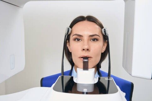
The scan is generally a quick, non-invasive imaging procedure. You will be positioned carefully while the scanner rotates around your head and captures multiple images.
Remaining still is important because movement can affect image quality. The captured images are then processed by computer software to create a three-dimensional view of the area being examined.
There is generally no recovery period following the scan itself. Your dentist can review the images and explain the findings during your consultation.
CBCT and Treatment Decisions
A CBCT scan is one part of the diagnostic process. The dentist considers the imaging findings together with your symptoms, examination, dental history, and treatment goals.
The scan may confirm information already suspected during an examination or reveal additional anatomical details that influence the treatment plan.
Importantly, advanced imaging is not automatically required for every dental problem. Your dentist determines whether the additional information provided by CBCT is clinically justified.
When Should You Ask About CBCT Imaging?
If you have been advised to undergo complex dental treatment, you can ask your dentist whether three-dimensional imaging would be useful for your case.
CBCT may be particularly relevant when conventional X-rays have not provided sufficient information or when the exact position of teeth, roots, bone, or surrounding structures needs to be evaluated.
Your dentist can explain the purpose of the scan, what it is expected to show, and how the results may influence your treatment.
Final Thoughts
Detailed imaging can be valuable when dental conditions involve complex anatomy or require careful treatment planning. When clinically appropriate, a Dental CBCT Scan can provide three-dimensional information that supports diagnosis and helps dental professionals better understand the structures involved in a patient's oral care.
Replies