Acetylcholinesterase and Its Biological Effects
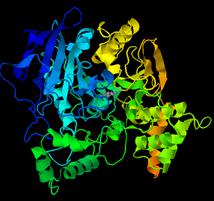
Acetylcholinesterase, abbreviated as AchE, is a key enzyme in biological nerve conduction. In cholinergic synapses, this enzyme can degrade acetylcholine, stop the excitatory effect of neurotransmitter on postsynaptic membrane, and ensure that nerve signals are in Normal transmission in organisms. It has the activity of carboxypeptidase and aminopeptidase. Acetylcholinesterase is involved in the development and maturation of cells and can promote neuron development and nerve regeneration.
Acetylcholinesterase, an essential enzyme with high activity and selective hydrolysis of Ach, can hydrolyze acetylcholine (ACh) into choline and acetic acid. The type I (true choli-neesterase) of cholinesterase has high substrate specificity because it only decomposes a narrow range of substrates centered on acetylcholine, so it is called this way.
Acetylcholinesterase is a key enzyme in biological nerve conduction. In cholinergic synapses, this enzyme can degrade acetylcholine, stop the excitatory effect of neurotransmitter on postsynaptic membrane, and ensure the normality of nerve signals in the organism transfer.
Metabolic mechanism
Studies by Chubbe et al. proved that AchE has carboxypeptidase and aminopeptidase activities. In vitro, AchE can hydrolyze enkephalin (Enk) and substance P (SP), but not somatostatin (Som) and vasopressin (VSP). Further studies have proved that as a peptidase, AchE has different active sites for hydrolyzing peptides and esterases. It is worth noting that many non-cholinergic neurons in the nervous system, which contain a large amount of AchE, also contain various neuropeptides. For example, SP cells in the dorsal root ganglion of the spinal cord are strongly positive for AchE.
Recent studies have shown that highly purified AchE from electric eel organs or bovine serum has protease-like or exonuclease activity. For serum proteins, AchE can eliminate the C-terminal residues. In addition, the protease-like action of AchE is also supported by molecular biology evidence. Amino acid analysis shows that the AchE protein molecule is similar to the amino acid sequence of protease-like endonuclease and serum carboxypeptidase. Within 36 residues of their C-terminal, 40% of the amino acid sequence is identical to the active fragment of the protease.
Biological effect
Release
Dendritic/cell body release is a special form of neurosecretion. The substantia nigra dopamine neurons are non-cholinergic and seem to rarely receive cholinergic afferent projections, but the substantia nigra cells contain a large amount of AchE. Studies have found that AchE in the brain can be membrane-bound and non-membrane-bound (soluble). The dendrites or cell bodies of substantia nigra dopaminergic neurons can secrete AchE (soluble) into the extracellular fluid. Among them, it is called the AchE dendritic release phenomenon. Obviously, the dendritic release of AchE has nothing to do with the release of Ach. Because the application of cholinergic blockers or antagonists does not affect the dendritic release of AchE. At the same time, the distribution of AchE in the ventricle and the content of cerebrospinal fluid are inconsistent with Ach. Since electron microscopy observations do not find the presence of dendritic synapses in the substantia nigra, the substances released by the dendrites or cell bodies in the substantia nigra can be relatively free diffusion. Therefore, AchE may play a non-synaptic regulatory role in the substantia nigra to regulate the sensitivity of the cell bodies of substantia nigra-striatal projection neurons to afferent signals from their distal dendrites.
Physiological behavior effect
Studies have found that extracellular AchE can change the electrical activity of substantia nigra dopamine neurons and animal movement behavior. When applied in large doses, AchE inhibits the spontaneous discharge of substantia nigra dopaminergic cells, but at physiological doses, it can increase the discharge frequency of substantia nigra cells. Creenfield found that the addition of AchE to the perfusion could cause significant hyperpolarization of the substantia nigra cell membrane with a decrease in membrane input resistance through in vitro cultured intracellular recording. The irreversible cholinesterase inhibitor Soman was used in advance. Processing AchE and then adding it to brain slice culture medium cannot change the influence of AchE on the electrical properties of the membrane. The above experiments show that the electrical properties of AchE on the membranes of substantia nigra dopaminergic neurons have nothing to do with the hydrolysis of Ach by AchE, and it has a high degree of structural specificity.
AchE was directly injected into the substantia nigra of rats with microelectrodes, which caused changes in the rats' motor behaviors, which were manifested as slow behavior and stupor. The behavioral changes of rats after an injection of AchE can last a long time, and the mechanism is not yet clear. It is speculated that under physiological conditions, after AchE is released from the dendrites of substantia nigra dopaminergic cells, it can have a certain impact on the physiological activities of itself and neighboring cells. This may be a new local connection between neurons or self-feedback. The way of adjustment.
Neurodevelopmental effects
More and more studies have suggested that AchE may be related to cell development and maturation. The evidence is: (1) Many embryonic tissues are rich in AchE; (2) AchE in serum of various primary tumor tissues and some cancer patients (3) AchE is closely related to the development of the nervous system.
Biochemical analysis and histochemical studies suggest that during development, AchE is related to the proliferation of nerve cells and the growth of neuronal processes. Because AchE appears during development, it appears in the same phase of development of different types of cells, and AchE appears the period coincides with the early stages of cell proliferation, migration, protrusion growth time, and synapse establishment. In conclusion, despite the lack of direct evidence so far, more and more research results support the speculation that AchE may act as a specific neurosecretory protein in the process of neurodevelopment, either in the cell or secreted outside the cell Regulates the proliferation of neurons and the growth of processes.
Nerve regeneration
Nerve injury and regeneration are very complex processes, which are regulated and influenced by many internal and external factors. It is known that the activity of AchE in neuron cell bodies and proximal axons is enhanced in the early stage of nerve injury. In the study of the relationship between AchE and nerve regeneration, some people used the irreversible AchE inhibitor DFP to chronically treat rats with sciatic nerve injury, which significantly weakened the ability of nerve regeneration. The exact mechanism by which DFP affects nerve regeneration is still unclear. One possibility is that AchE can promote nerve regeneration, and DFP, as an organophosphate poison, irreversibly combines with AchE protein molecules, which prevents AchE from acting in nerve regeneration.
As we all know, peripheral nerves have stronger regeneration ability than central nerves. It has been discovered in medicine that central nervous tissues also have a certain ability to regenerate, especially those older fibers in germline, such as the hippocampal pathway, the hypothalamic-pituitary fiber, and the central monoaminergic pathway. It is worth noting that these nerve fibers or cell bodies contain high levels of AchE. Combining AchE with neuron development, neurite outgrowth and its relationship with nerve regeneration, it is proposed that AchE not only participates in the transmission of cholinergic neurotransmitters, but also has neurotrophic factor-like effects that regulate and promote nerve tissue development and nerve regeneration.
Acetylcholinesterase related Assay Kit
Creative BioMart specializes in providing cost-effective assay kits and custom services to both academia and biotech/pharmaceutical industries in the study of enzyme activity, protein biology, and small molecule detection. For Acetylcholinesterase Assay Kit, Creative Biomart offers Acetylcholinesterase Activity Colorimetric Assay Kit, Acetylcholinesterase Assay Kit,Acetylcholinesterase Green Fluorescence Assay Kit, Acetylcholinesterase Red Fluorescence Assay Kit etc.
Replies